
A sample can preserve what a cell looks like while losing the chance to study what it does. That gap is the target of a new engineering approach from MIT and Johns Hopkins researchers.
In work described by MIT on 24 September, a handheld microfluidic device collected living cells from selected areas of freshly removed tissue. It directs fluid across a small surface region, using the resulting shear force to release cells for collection. Researchers recovered cells that could be grown for further study, including as organoids. The system is described in the journal Device. MIT’s research report
Preserving location and behaviour
The research focused on sampling removed fallopian-tube tissue, motivated by the need to study early changes associated with ovarian cancer. Its promise lies in obtaining living cells from a specific location while preserving surrounding tissue for pathology.
The crucial boundary is the experimental setting: this is a tissue-sampling research tool. The reported work does not establish a screening test that can detect ovarian cancer in otherwise healthy people, or a reduction in cancer mortality. Study context and limitations
The engineering question behind the headline
Our assessment is that the most compelling aspect is the potential connection between a cell’s origin and its later behaviour in the laboratory. That could help researchers frame better experiments. It does not automatically make the resulting experiment clinically predictive.
Several questions would matter in further validation. Does collection favour some cell types over others? How consistently can different operators recover comparable samples? Does growth in the laboratory alter the properties researchers are trying to investigate?
Those questions are especially relevant if a future application involves comparing responses to possible treatments. A result in a dish would still need evidence linking it to what happens in a patient.
There is also a workflow test. A new device has to fit the handling, timing and interpretation of real samples. An elegant collection method will be more useful if it produces reproducible material without creating uncertainty about the tissue that remains.
This is the kind of advance that can matter before it becomes a clinical product. Better access to useful samples can open experiments that were previously difficult to perform. The next milestones should show how reliably that access improves research, and only then whether it can support a validated diagnostic application.
Sources
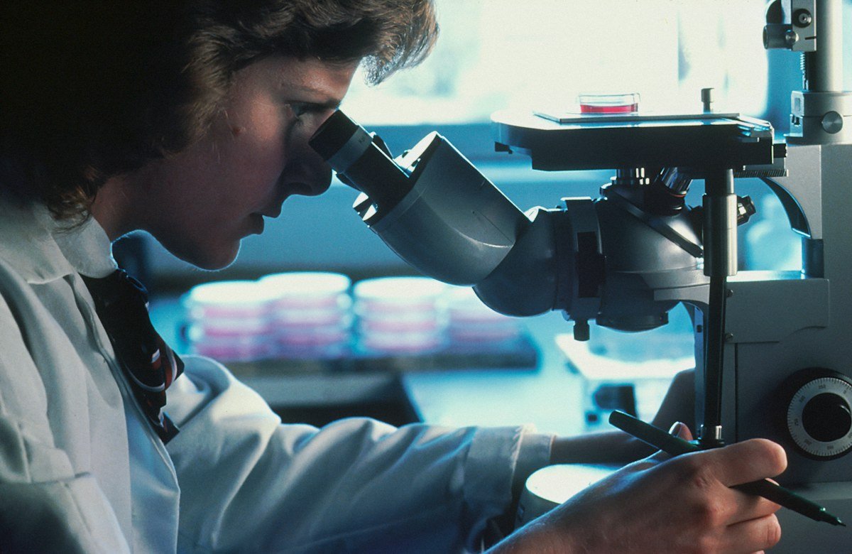
Featured image: Representative laboratory photograph, not the MIT cell-collection device. Credit: National Cancer Institute.
Leave a Reply