
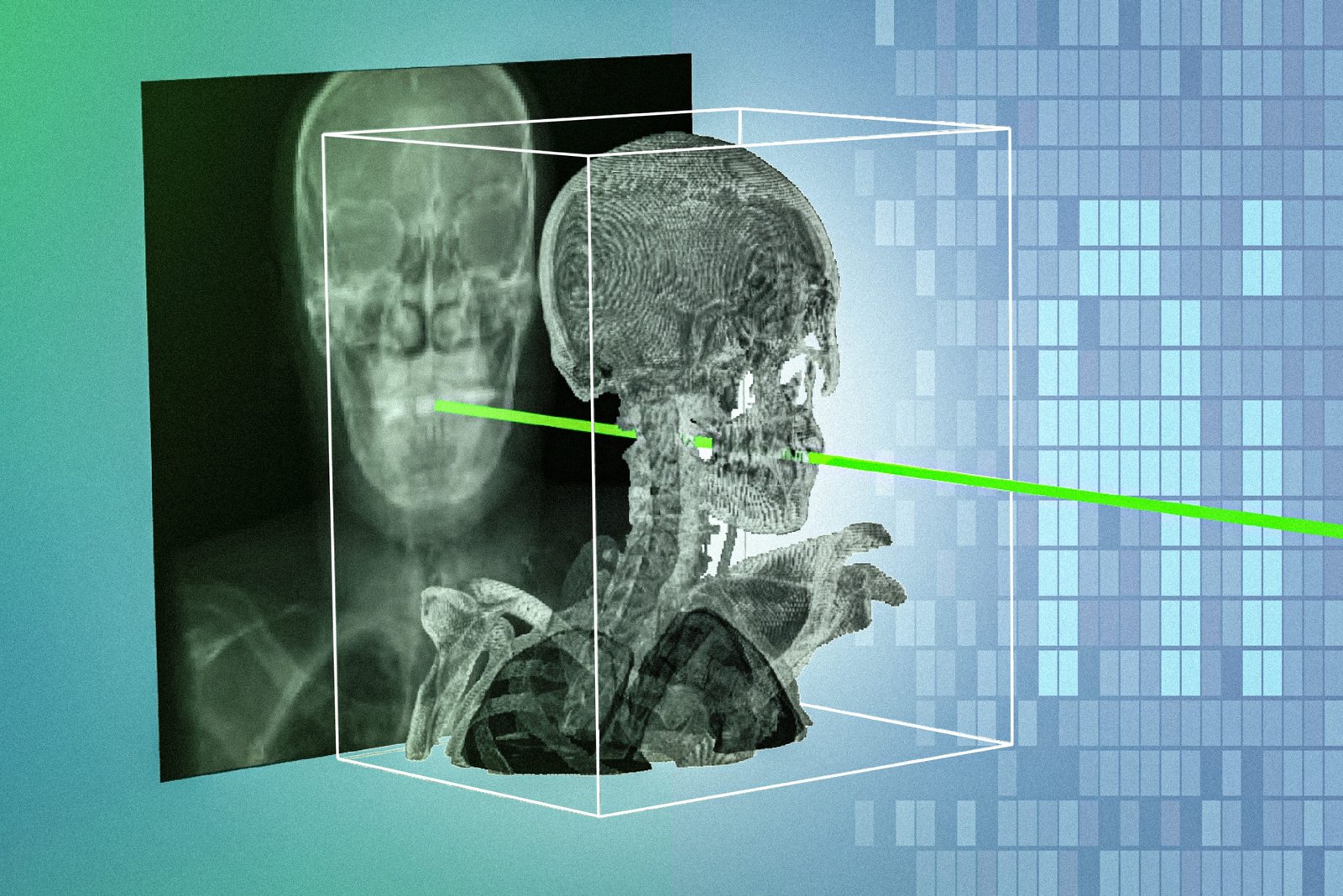
Imagine navigating a building with a detailed three-dimensional model, while the view in front of you is a flat photograph taken from an unfamiliar angle. Both show the same place. Working out exactly how they fit together is the difficult part.
Surgeons can face a related challenge when matching X-rays taken during a procedure with a patient’s earlier CT or MRI scan. A new AI method, called xvr, is designed to make that alignment faster and more dependable. The research was published in Nature on 16 September 2026. Nature paper
Its central idea is unusually personal: adapt the model to the particular patient whose anatomy it needs to understand.
Five minutes of adaptation
Xvr stands for X-ray volume registration. Registration means matching images so that corresponding structures line up, even when the images were captured in different ways or from different positions.
The system uses a patient’s existing three-dimensional scan to simulate X-rays from many angles. A pretrained model can then adapt to that patient in about five minutes. The team reports that subsequent matching takes seconds, with submillimetre precision in its evaluations. MIT’s explanation
Five minutes refers to adaptation of an already trained system. It is not the total time needed to invent, train and validate the underlying technology.
The research combines neural networks with further mathematical refinement of the image alignment. The paper describes its scope as rigid registration: matching position and orientation, rather than solving every way that living tissue can bend or change shape. Nature
A map has to remain trustworthy
The team evaluated data covering different body regions and patients from five hospitals. MIT says further work includes additional reliability studies and handling more complex situations such as moving body parts. MIT
Those next steps matter because an accurate-looking overlay can be persuasive. A tool intended to support navigation must also make it possible to recognise when the match is unreliable.
Our assessment is that the reported alignment results justify interest, while leaving a separate clinical question open: whether using the system improves outcomes during real procedures. This study is not evidence that patients already experience fewer complications, and it is not an announcement of regulatory approval.
Other researchers can inspect the approach
The team has released the xvr software, along with pretrained models and material for reproducing reported experiments. Its public repository describes tools for training models and registering X-rays to CT or MRI data. xvr research software
That gives the wider research community something concrete to examine. Reproducible software can help reveal which parts of a result survive changes in data, equipment and operating conditions. It does not, on its own, make a medical product ready for clinical use.
The appeal of xvr is its focus on a specific, difficult task. It aims to help clinicians connect information they already have, using the individual patient as the starting point.
If that approach proves reliable in practice, one of AI’s most useful contributions to surgery could be a clearer answer to a very practical problem: knowing precisely how the view on a screen relates to the body in front of the surgeon.
Related reading: Targeted MRI research for detecting lung cancer.
Illustration of matching X-rays with three-dimensional medical scans. Credit: Courtesy of the researchers; MIT News. This is a research illustration.
Leave a Reply