
Ageing research has a measurement problem. Before scientists can judge whether an intervention changes troublesome cells, they need a reliable way to identify those cells in the first place.
RamanOmics, highlighted by MIT on 21 September 2026, aims to make that job more precise. It combines light-based chemical measurements with gene-activity information and machine learning to identify patterns associated with senescent cells. The reported work involved mouse tissue; adaptation to human samples is still underway. MIT says the study was published in Nature Aging. MIT announcement and journal link
Senescent cells have stopped dividing but remain present. They are sometimes called “zombie cells”, an attention-grabbing nickname for a biological state that is more complicated than simply being harmful.
A barcode made from chemistry
Raman imaging reads how light interacts with molecules. Different chemical bonds contribute different features to the resulting signal. In this work, those measurements were combined with information about which genes were active and where cells sat within tissue.
The authors’ earlier, openly available preprint describes that combined approach in mouse lung and skin. Machine learning helped assemble several signals into a barcode associated with senescence, rather than relying entirely on one marker. It also explored how the patterns changed in a wound-healing model. Earlier research version, indexed as a preprint
“Barcode” is a metaphor here. Nothing is printed on the cell. Researchers are looking for a recognisable pattern in data.
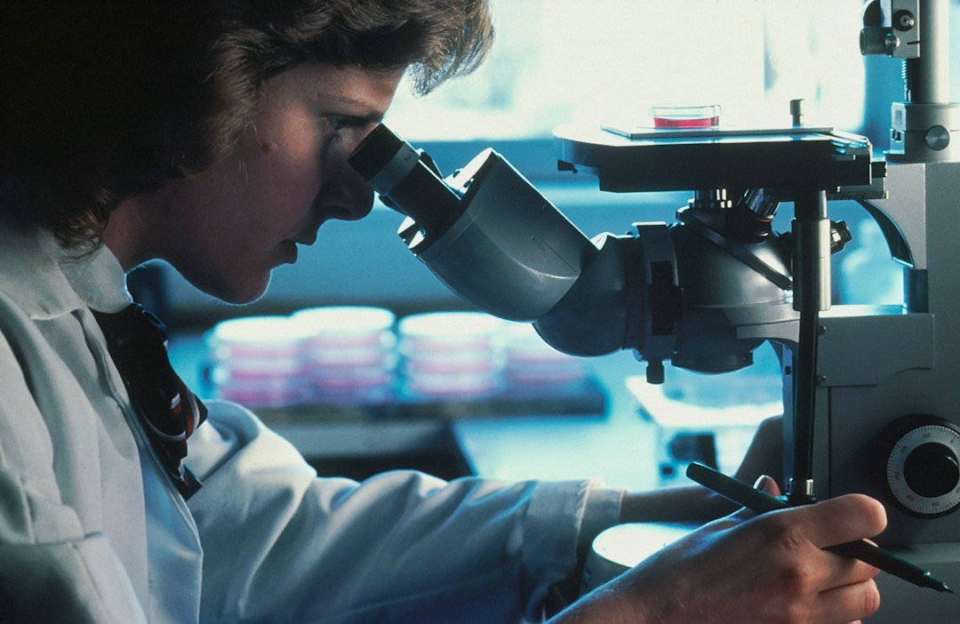
The distinction between developing the barcode and eventually reading it is useful. Detailed molecular analyses help establish what a light-based signal means. The longer-term goal is to recognise informative patterns with imaging that preserves the specimen, without having to repeat every destructive analysis.
The scanner is still slow—and the biology is not simple
According to MIT, analysing a tissue area of roughly one square millimetre currently takes about 30 hours. The team is working on faster imaging. That is a substantial practical gap between a research demonstration and anything resembling a routine examination. Current limitations
The word senescence also does not identify a universal enemy. MIT notes that the process contributes to normal development and tissue repair as well as age-related disease. A future system would need to help interpret cells in context, not merely count every positive signal as something to eliminate.
Our assessment is that this is why measurement deserves attention alongside proposed longevity treatments. A more informative test could help researchers ask whether an intervention changed the intended cell population, in the intended tissue, at the intended time.
Better evidence comes before an anti-ageing claim
This study did not show longer human lifespan, reverse ageing in patients or validate a consumer biological-age test. It developed and investigated a way of characterising cells.
The next questions concern reproducibility across laboratories, performance in human tissues and whether faster readings preserve useful accuracy. Even successful identification would leave the separate problem of deciding what, if anything, should be done about those cells.
For readers following experimental models built from human tissue, the shared lesson is that better research tools can be valuable before they become treatments.
The promising part of RamanOmics is its potential to make a difficult biological target easier to study. Its first contribution is to the quality of the questions scientists can answer.
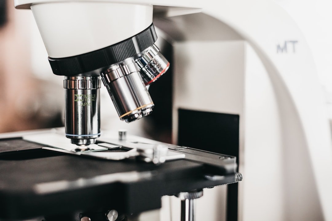
Featured image: Representative laboratory microscope; this is not the RamanOmics instrument used in the study. Image: Logan Gutierrez / Unsplash.
Leave a Reply